logo
  Home | Contact us | Browse | Quick Search by UniProtKB ID, Keyword, PDBID

Menu:

Latest news:

Sep. 10, 2014:
A total of 174 experimentally verified S-nitrosylation sites on 94 S-nitrosylated proteins from individualized human colorectal cancer tissues using a label-free quantitation strategy.

Read more...


Protein Name: E3 ubiquitin-protein ligase Mdm2

UniprotKB/SwissProt ID: MDM2_HUMAN (Q00987)

Gene Name: MDM2

Organism: Homo sapiens (Human).

Function: E3 ubiquitin-protein ligase that mediates ubiquitination of p53/TP53, leading to its degradation by the proteasome. Inhibits p53/TP53- and p73/TP73-mediated cell cycle arrest and apoptosis by binding its transcriptional activation domain. Also acts as a ubiquitin ligase E3 toward itself and ARRB1. Permits the nuclear export of p53/TP53. Promotes proteasome-dependent ubiquitin-independent degradation of retinoblastoma RB1 protein. Inhibits DAXX-mediated apoptosis by inducing its ubiquitination and degradation. Component of the TRIM28/KAP1-MDM2-p53/TP53 complex involved in stabilizing p53/TP53. Also component of the TRIM28/KAP1-ERBB4-MDM2 complex which links growth factor and DNA damage response pathways. Mediates ubiquitination and subsequent proteasome degradation of DYRK2 in nucleus. Ubiquitinates IGF1R and SNAI1 and promotes them to proteasomal degradation.

Other Modifications: View all modification sites in dbPTM

Protein Subcellular Localization: Nucleus, nucleoplasm. Cytoplasm. Nucleus, nucleolus. Note=Expressed predominantly in the nucleoplasm. Interaction with ARF(P14) results in the localization of both proteins to the nucleolus. The nucleolar localization signals in both ARF(P14) and MDM2 ma

PDB :
( If your security settings prevent Jmol from running, please register http://140.138.144.145/ as a safe location in your Java settings. )

Protein disease:
Disease database Database Entry Disease information
HPRD01272Accelerated tumor formation, susceptibility to
Network with metabolic pathway:
Kegg map ID Pathway Link
map05219Bladder cancer
map04115p53 signaling pathway
map05214Glioma
map05215Prostate cancer
map05218Melanoma
map05220Chronic myeloid leukemia
map04110Cell cycle
map04151PI3K-Akt signaling pathway
map05169Epstein-Barr virus infection
map05200Pathways in cancer
map05202Transcriptional misregulation in cancer
map05203Viral carcinogenesis
map04120Ubiquitin mediated proteolysis
map04144Endocytosis
Graphical Visualization of S-nitrosylation Sites:
Overview of Protein S-nitrosylation Sites with Functional and Structural Information
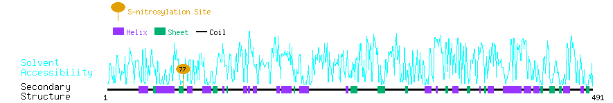
InterPro ID Domain
IPR001841
IPR001876
IPR003121

3D Structure Databases:
3D structure databases
EntryMethodResolution (A)ChainPositionsView
1RV1 X-ray 2.30 A A/B/C25-109Link
1T4E X-ray 2.60 A A/B17-111Link
1T4F X-ray 1.90 A M17-125Link
1YCR X-ray 2.60 A A17-125Link
1Z1M NMR - A1-118Link
2AXI X-ray 1.40 A A17-125Link
2C6A NMR - A290-335Link
2C6B NMR - A290-335Link
2F1Y X-ray 1.70 A A-Link
2FOP X-ray 2.10 A B145-150Link
2GV2 X-ray 1.80 A A17-125Link
2HDP NMR - A/B429-491Link
2LZG NMR - A1-125Link
2M86 NMR - B17-125Link
2VJE X-ray 2.20 A A/C428-491Link
2VJF X-ray 2.30 A A/C428-491Link
3EQS X-ray 1.65 A A25-109Link
3G03 X-ray 1.80 A A/C18-125Link
3IUX X-ray 1.65 A A/C25-109Link
3IWY X-ray 1.93 A A/C25-109Link
3JZK X-ray 2.10 A A17-111Link
3JZR X-ray 2.10 A A17-125Link
3JZS X-ray 1.78 A A24-109Link
3LBK X-ray 2.30 A A18-111Link
3LBL X-ray 1.60 A A/C/E18-111Link
3LNJ X-ray 2.40 A A/C/E25-109Link
3LNZ X-ray 1.95 A A/C/E/G/I/K/M/O25-109Link
3TJ2 X-ray 2.10 A A/C18-111Link
3TPX X-ray 1.80 A A/C/E25-109Link
3TU1 X-ray 1.60 A A18-125Link
3V3B X-ray 2.00 A A/B24-110Link
3VBG X-ray 2.80 A A/B/C/D25-109Link
3VZV X-ray 2.80 A A/B25-109Link
3W69 X-ray 1.90 A A/B25-109Link
4DIJ X-ray 1.90 A A/B17-111Link
4ERE X-ray 1.80 A A/B17-111Link
4ERF X-ray 2.00 A A/C/E17-111Link
4HBM X-ray 1.90 A A/B/C/D/E/F/G/H6-125Link
4HFZ X-ray 2.69 A A/C17-125Link
4HG7 X-ray 1.60 A A17-108Link
4JV7 X-ray 2.20 A A18-111Link
4JV9 X-ray 2.50 A A18-111Link
4JVE X-ray 2.30 A A18-111Link
4JVR X-ray 1.70 A A/C/E18-111Link
4JWR X-ray 2.35 A A/B/C17-111Link
4MDN X-ray 1.90 A A18-110Link
4MDQ X-ray 2.12 A A25-110Link
4OAS X-ray 1.70 A A/C/E17-111Link

The S-nitrosylation sites of MDM2_HUMAN

No. Position S-nitrosylated Peptide Secondary Structure of S-nitrosylated Peptide Solvent Accessibility of nitrosylated Site Substrate Motifs PubMed ID Experiment
177YDEKQQHIVY C SNDLLGDLFG HHCCCCEEEE E CCCHHHHHHC 1.81%HC0122178444-
277YDEKQQHIVY C SNDLLGDLFG HHCCCCEEEE E CCCHHHHHHC 1.81%HC0115688001-