logo
  Home | Contact us | Browse | Quick Search by UniProtKB ID, Keyword, PDBID

Menu:

Latest news:

Sep. 10, 2014:
A total of 174 experimentally verified S-nitrosylation sites on 94 S-nitrosylated proteins from individualized human colorectal cancer tissues using a label-free quantitation strategy.

Read more...


Protein Name: Transforming protein RhoA

UniprotKB/SwissProt ID: RHOA_MOUSE (Q9QUI0)

Gene Name: Rhoa

Synonyms: Arha, Arha2

Organism: Mus musculus (Mouse).

Function: Regulates a signal transduction pathway linking plasma membrane receptors to the assembly of focal adhesions and actin stress fibers. Involved in a microtubule-dependent signal that is required for the myosin contractile ring formation during cell cycle cytokinesis. Plays an essential role in cleavage furrow formation. May be an activator of PLCE1. Activated by ARHGEF2, which promotes the exchange of GDP for GTP. Essential for the SPATA13-mediated regulation of cell migration and adhesion assembly and disassembly. The MEMO1-RHOA-DIAPH1 signaling pathway plays an important role in ERBB2-dependent stabilization of microtubules at the cell cortex. It controls the localization of APC and CLASP2 to the cell membrane, via the regulation of GSK3B activity. In turn, membrane-bound APC allows the localization of the MACF1 to the cell membrane, which is required for microtubule capture and stabilization (By similarity). Required for the apical junction formation of keratinocyte cell-cell adhesion.

Other Modifications: View all modification sites in dbPTM

Protein Subcellular Localization: Cell membrane; Lipid-anchor; Cytoplasmic side (By similarity). Cytoplasm, cytoskeleton (By similarity). Cleavage furrow (By similarity). Cytoplasm, cell cortex (By similarity). Midbody (By similarity). Cell projection, lamellipodium. Note=Translocates to

Network with metabolic pathway:
Kegg map ID Pathway Link
map04360Axon guidance
map04520Adherens junction
map04660T cell receptor signaling pathway
map04670Leukocyte transendothelial migration
map04722Neurotrophin signaling pathway
map04972Pancreatic secretion
map05100Bacterial invasion of epithelial cells
map05133Pertussis
map05210Colorectal cancer
map04062Chemokine signaling pathway
map04270Vascular smooth muscle contraction
map04310Wnt signaling pathway
map04350TGF-beta signaling pathway
map04510Focal adhesion
map04530Tight junction
map04810Regulation of actin cytoskeleton
map05152Tuberculosis
map05200Pathways in cancer
map05203Viral carcinogenesis
map04144Endocytosis
Graphical Visualization of S-nitrosylation Sites:
Overview of Protein S-nitrosylation Sites with Functional and Structural Information
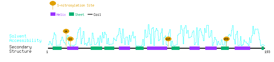
InterPro ID Domain
IPR001806
IPR003578
IPR005225
IPR013753

3D Structure Databases:
3D structure databases
EntryMethodResolution (A)ChainPositionsView
4F38 X-ray 2.80 A A1-191Link

The S-nitrosylation sites of RHOA_MOUSE

No. Position S-nitrosylated Peptide Secondary Structure of S-nitrosylated Peptide Solvent Accessibility of nitrosylated Site Substrate Motifs PubMed ID Experiment
116KKLVIVGDGA C GKTCLLIVFS EEEEEEECCC C CHHHHHHHHH 7.60%MC0621278135in vitro
220IVGDGACGKT C LLIVFSKDQF EEECCCCCHH H HHHHHHHCCC 3.11%MC0522865876in vivo
3107EKWTPEVKHF C PNVPIILVGN HHHHHHHHHH C CCCEEEEEEE 2.10%MC0821278135in vitro
4107EKWTPEVKHF C PNVPIILVGN HHHHHHHHHH C CCCEEEEEEE 2.10%MC0822865876in vivo
5159NRIGAFGYME C SAKTKDGVRE HHCCCCEEEE E EECCCCCHHH 2.46%MC0321278135in vitro
6159NRIGAFGYME C SAKTKDGVRE HHCCCCEEEE E EECCCCCHHH 2.46%MC0322865876in vivo